Working with complex, multi-system cases has kept me busy trying to explain how each symptom can be happening. So often the picture looks the same: a long list of symptoms that don't seem connected, conventional labs that come back normal (or normal apart from cholesterol and maybe slightly raised liver enzymes), and a body that still feels like nothing is working the way it should. These are the patterns and explanations that have made sense to me and to my clients, and I hope they help you connect a few dots too.
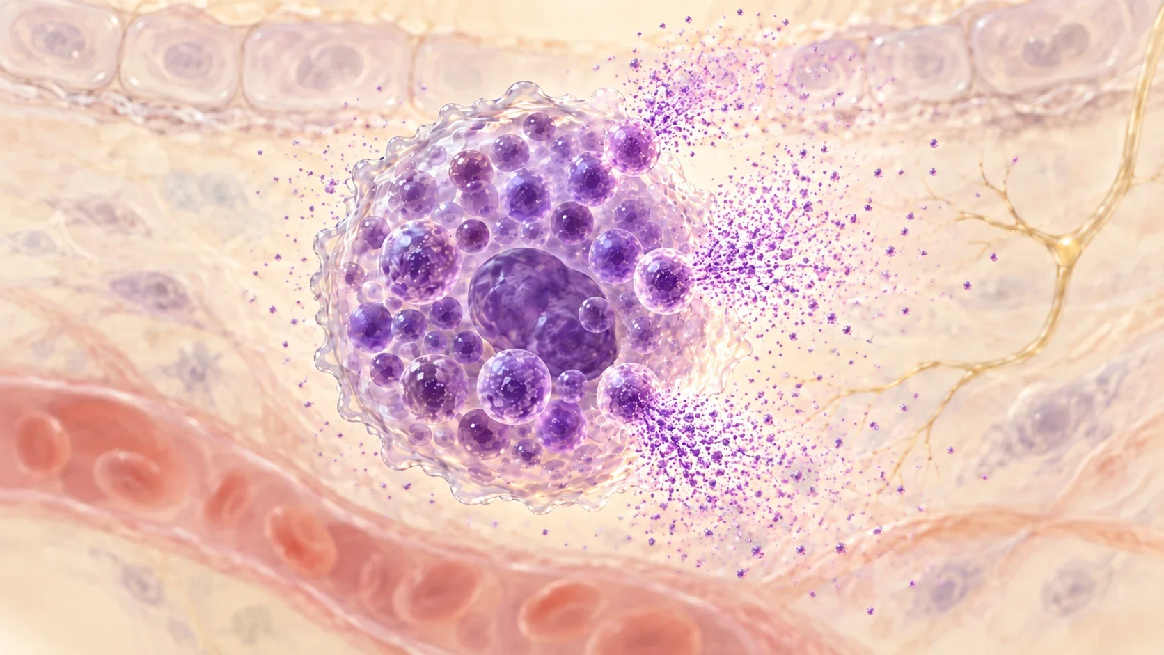
Every tissue that touches the outside world — skin, gut, airways, and the lining of every blood vessel — is guarded by mast cells. They are your sentinels: stationed at the borders, deciding in seconds whether something is a threat, and calling in help when it is. When they become hyperreactive, it is because the threats have been too many, too constant, or never fully resolved — so they stay on guard. The reactivity is protection that has not been able to stand down.
Histamine is only one of hundreds of signals a mast cell can release — a recent compilation of the human literature identified at least 390 (Molderings & Afrin, 2023). That is why "it's not allergies" and "I react to everything" can both be true.
Does this sound like you?
No one has all of these, and none of them alone means mast cell activation. The pattern that suggests it is symptoms in several unrelated systems at once, that come and go, and that don't line up with any single diagnosis.
Skin
- Flushing, warmth in the face, neck or chest
- Hives, welts, itching without a rash
- Skin that writes when you scratch it
- Rashes, eczema, burning or crawling sensations
- Easy bruising, slow healing
- Swelling that moves around; brittle or ridged nails
- Reacting to fabrics, detergents, fragrance or skincare
Gut and mouth
- Bloating, cramping, nausea, reflux
- Alternating loose and constipated
- Reacting to foods you used to tolerate, and a shrinking safe-food list
- Early fullness, appetite and weight changes
- Tingling, itchy or swollen lips, tongue or mouth, especially after eating
- Mouth ulcers or white patches on the tongue
- Burning mouth or tongue
- Metallic taste or other changes in taste
- Sensitive teeth or gums
- Difficulty swallowing
Heart and circulation
- Racing heart, palpitations, chest tightness
- Lightheaded on standing, near-fainting
- Blood pressure that runs low, runs high, or swings between the two, including sudden, very high spikes during a flare
- Cold hands and feet, poor circulation
- Swelling and puffiness that come and go
Brain and nerves
- Brain fog, word-finding trouble, poor concentration
- Memory and attention problems; feeling detached or unreal (depersonalization)
- Anxiety that arrives out of nowhere, or a sense of dread
- Insomnia, waking in the night, unrefreshing sleep, vivid dreams or night terrors
- Headaches or migraines
- Sensitivity to light, sound and smell
- Tingling or numbness, including in the face, that comes and goes; internal buzzing or tremor
- Dizziness, vertigo
- Low mood, irritability
Breathing and airways
- Air hunger, sighing, can't get a full breath
- Throat tightness or a lump-in-throat feeling
- Dry cough, hoarseness, wheeze; recurring bronchitis
- Excess mucus or phlegm and throat clearing that keeps coming back, especially after eating or near a trigger
Eyes, ears and nose
- Red or bloodshot, itchy, burning, gritty or grainy eyes; eyelid twitching
- Watery or dry eyes; puffy or swollen eyelids
- Dark under-eye circles
- Blurry vision or trouble focusing that comes and goes
- Itchy or full ears, pressure, tinnitus, hearing that fluctuates
- Chronic congestion, runny nose, sneezing fits, post-nasal drip, sinusitis
- Nosebleeds, sores inside the nose; changes in sense of smell
Whole body
- Fatigue that rest doesn't fix; crashing after exertion
- Temperature swings, sweats, feeling cold much of the time
- Muscle and joint pain, or pain and swelling that moves from joint to joint; joints that are unusually bendy, or that click, subluxate or feel unstable
- A heavy or painful head and neck, headaches at the base of the skull, dizziness or visual trouble that worsens the longer you are upright
- Bone pain, bone density loss
- Stretchy or velvety skin, scars that widen, slow wound healing
- Bladder frequency, urgency, pain or not fully emptying, with no infection found
- Cycle-linked flares, heavy or painful periods; fertility difficulties
- Vulvar or vaginal itching or pain, painful intercourse, low libido; prostate inflammation
- Blood sugar swings; raised cholesterol or triglycerides
- Infections that linger or keep returning
- Tender lymph nodes that come and go
- Reacting to chemicals, fragrance, exhaust, new furnishings or building work
- Reacting to medications, supplements, or the fillers in them
What a mast cell is
A tissue-resident immune cell, born in the bone marrow, that moves into a tissue and lives there for months to years — packed with pre-loaded granules ready to fire.
Pre-loaded
- Granules hold histamine, tryptase, chymase, heparin and more — already made, released in seconds
- A second wave (lipid signals) is made within minutes
- A third wave (cytokines) over hours
Sentinel, defender, repair coordinator
- Sits at borders: next to blood vessels, nerves and epithelium
- Detects danger, opens the gates, calls in other immune cells — and kills some microbes directly
- Coordinates tissue repair; also senses hypoxia, pressure and vibration
- The goal is better regulation, not treating these cells as the enemy
Shaped by its neighborhood
- Skin, connective tissue and vessel wall mast cells carry a fuller set of enzymes — they do more tissue remodeling and more of the blood-vessel work
- Gut and airway lining mast cells carry a narrower set and shift with what is happening at that surface
- A mast cell in the gut can behave quite differently from one in the brain or lungs, shaped by the tissue around it — which is part of why this looks so different from person to person
- Numbers rise with chronic inflammation — the tissue "recruits" more
Where they live
Everywhere the body meets the world, and everywhere blood meets tissue. Their location explains why a flare can feel like it is "everywhere at once."
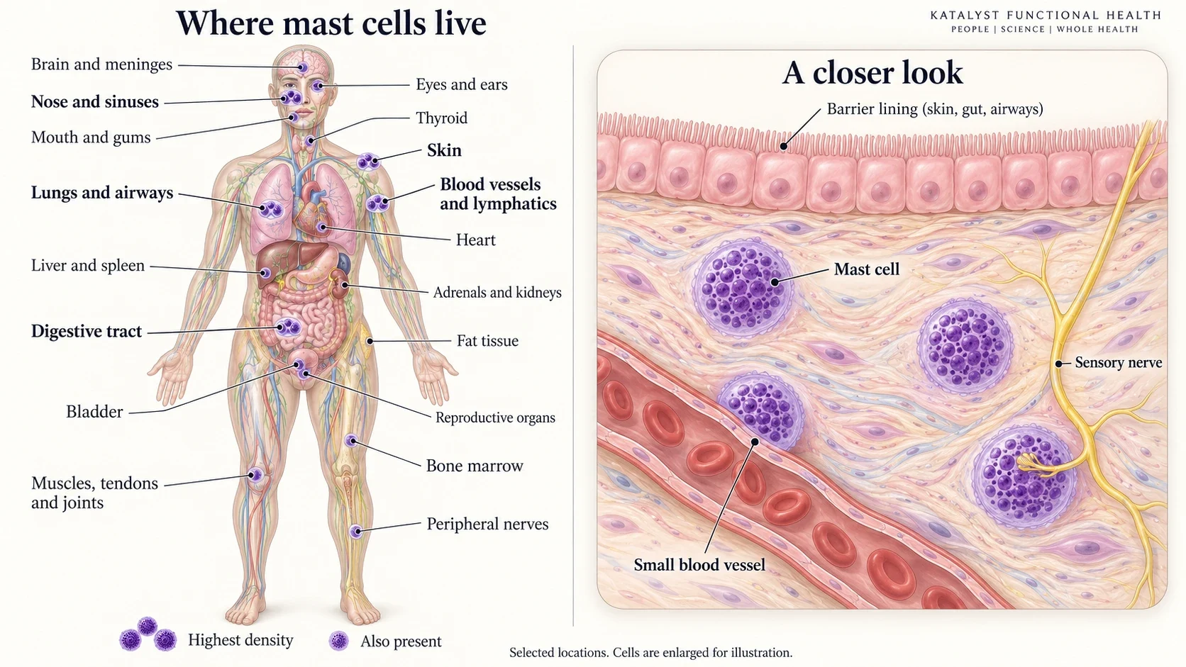
Highest density
- Skin (dermis)
- Nose, sinuses, airways, lungs
- Gut lining and around gut nerves
- Around nearly every blood vessel and lymphatic
Also present and clinically important
- Brain: meninges, thalamus, hypothalamus, near the amygdala and pineal; spinal cord and nerve ganglia
- Heart (beside its nerves), pericardium, carotid bodies, adrenals
- Eyes, ears, mouth, dental pulp, thyroid
- Liver, spleen, pancreas, kidneys, peritoneum
- Fat tissue — a major and often forgotten reservoir
- Muscle, fascia, tendons, joint lining (synovium)
- Bladder, uterus, ovaries, prostate, testes, breast, placenta
- Bone marrow and bone surfaces
- Closely associated with peripheral nerves — mast cells and nerve endings often sit in direct contact (Forsythe, 2019)
What one activated mast cell releases
Three waves, dozens of the most important signals shown here. Color shows the wave: pre-formed granule contents fire first, lipid messengers within minutes, cytokines and growth factors over hours.
Mast cells release selectively: a stress trigger, a bacterial trigger and an allergic trigger each produce a different mix (Theoharides et al., 2007) — which is why one person's flare is flushing and another's is fatigue and pain.
Felt in minutes
- Flushing, itch, hives, swelling — histamine, PAF
- Drop in blood pressure, racing heart — histamine, PGD2
- Wheeze, throat tightness, mucus — leukotrienes, PGD2
- Cramping, urgency, nausea — histamine, serotonin
Felt in hours
- Fatigue, achiness, "flu-like" — IL-1β, IL-6, TNF
- Brain fog, low mood, anxiety — IL-6, TNF, histamine on the brain
- Itch without hives — IL-31, tryptase on nerves
- Pain amplification — nerve growth factor, tryptase
Felt over months
- Tissue thickening and scarring — TGF-β1, chymase, tryptase, MMP-9
- Bone loss — histamine, heparin, IL-6
- Leaky gut, leaky vessels, leaky lymph — tryptase, VEGF, heparanase
- New vessel growth, remodeling — VEGF, FGF
What pushes the button
A mast cell carries many different "docking stations" on its surface. IgE allergy is only one of them. People with mast cell activation can react through true allergies and non-allergic triggers together, or through non-allergic triggers alone: plenty of people with strong mast cell reactivity have no allergies at all.
Microbial triggers common in chronic illness
- LPS (endotoxin) from gram-negative bacteria — gut, water-damaged buildings → TLR4
- Peptidoglycan and lipoteichoic acid from gram-positive bacteria, including Actinomycetes → TLR2 (Supajatura et al., 2002)
- β-glucans from mold cell walls, spores and spore fragments → Dectin-1 (Olynych et al., 2006)
- Oxalates often travel with fungal overgrowth — Aspergillus and Candida produce them, and the crystals irritate tissue mechanically, adding another local input on top of the fungal one
- Viral RNA/DNA → TLR3, TLR7, TLR9, from active and reactivated viruses such as EBV and HHV-6
- Spike protein — a strong driver since 2020. It acts on mast cells through ACE2 and TLR4 (Tsilioni & Theoharides, 2023), and spike-specific IgE has now been measured in patients (de la Poza et al., 2025), adding an allergy-type route on top. Spike can linger long after infection or vaccination: for months, and in some people for years. After infection it can hide in the gut lining, bone marrow and circulating immune cells, so the trigger keeps firing long after the acute illness has passed. The ongoing inflammation can also wake up dormant infections such as EBV and Lyme
- Bacterial and fungal proteases — from fungi such as Aspergillus, Alternaria, Penicillium and Candida, and bacteria such as Staphylococcus aureus, Pseudomonas aeruginosa, Porphyromonas gingivalis (gum disease) and Streptococcus. Fungal proteases can be inhaled from the environment or produced by fungi colonizing the gut, sinuses or skin. They cut and switch on PAR-2 receptors on mast cells and the tissue lining, release IL-33 from damaged tissue (Cayrol et al., 2018), and loosen barriers
- Borrelia (Lyme) and Bartonella, through their surface proteins. Both are more common than previously thought. They can cause chronic, subclinical or multi-symptom infection, with complex immune evasion, biofilms and dormant states that help them escape conventional detection. Bartonella lives in the vessel lining itself: it drives endothelial inflammation and switches on the low-oxygen alarm (HIF-1) and VEGF (Riess et al., 2004; Kempf et al., 2005), partly by using up oxygen inside the cells it infects, all of which push the mast cells sitting against those vessels
- Parasites — mast cells are a primary defense against parasites, so they respond powerfully: IgE-driven activation, direct antigen recognition, and IL-33 from the tissue damage the organism causes (Mukai et al., 2016). Where a parasite is found and treated, reactivity sometimes settles alongside it
- Complement fragments C3a and C5a — released whenever the immune system is fighting anything
Nerve, hormone and stress triggers
- CRH — the first stress hormone out of the brain — binds mast cells directly (CRH-R1) (Cao et al., 2005)
- Substance P, NGF, CGRP, VIP, PACAP released by irritated nerves → MRGPRX2 and neurokinin receptors (Green et al., 2019)
- Estrogen can enhance some responses, and when it dominates relative to progesterone it raises inflammatory cytokines and lowers the threshold for degranulation (Zaitsu et al., 2007); progesterone and testosterone have both calmed mast cells in lab studies (Vasiadi et al., 2006, for progesterone). Effects depend on tissue, so cycle patterns are assessed individually
- Thyroid hormone, cortisol imbalance and adrenaline shift the threshold rather than trigger outright. Low thyroid slows cellular energy production, and less energy means less capacity for detoxification and repair — one plausible route to a lower bar for activation
- Poor sleep and circadian disruption lower the threshold (Nakamura et al., 2014)
Self-amplifying: the cell re-triggers itself
- Histamine acts back on the mast cell (H1, H4) and on neighbors
- Leukotrienes and PGD2 bind receptors on mast cells and pull more in
- ATP spilled by injured cells → P2X7 receptor
- IL-33 (alarmin from damaged epithelium) → ST2 — one of the strongest amplifiers (Allakhverdi et al., 2007)
- Stem cell factor (SCF/KIT) keeps them alive and multiplying
- DAMPs from stressed cells: HMGB1, uric acid, mitochondrial DNA
Physical and chemical triggers
- Heat, cold, sun, pressure, friction, vibration (vibration plates, ultrasound, loud bass)
- Barometric pressure and weather changes — a very common report, and one reason flares track storm fronts
- Exertion and heat from exercise; sudden temperature shifts, hot showers
- Hypoxia and adenosine — low oxygen delivery is itself a trigger
- Alcohol and aldehydes, sulfites, food dyes, salicylates in sensitive people
- Ammonia and cationic amines — in laboratory work, ammonia alkalinizes the cell and can trigger histamine release on its own; cationic amines act through MRGPRX2
- Some medications, acting through MRGPRX2 (McNeil et al., 2015) — the same receptor substance P uses
- Heavy metals, particularly mercury, and mycotoxins can activate mast cells directly (Kempuraj et al., 2010, for mercury); pesticides and other toxicants act more through oxidative stress and damage to the surrounding tissue
- Electromagnetic fields — in human studies, phone exposure enlarged allergen-triggered skin reactions and raised substance P, pointing at the mast cell itself (Kimata, 2002); the proposed mechanism is opening of voltage-gated calcium channels
Classic allergy vs. mast cell activation
Allergy is one key in one lock. Mast cell activation is a cell with every door propped open and the threshold set too low. Normal allergy tests alongside dramatic reactions is a common picture, not a contradiction. The two aren't mutually exclusive either: someone can have genuine IgE allergies and broader mast cell reactivity at the same time, and each adds to the other's load. The description people most often give is that their body suddenly became reactive to everything, and stopped being predictable.
IgE allergy
- Specific and identifiable: tree, grass and weed pollens; dust mite; cockroach; cat, dog and other animal dander; mold spores; latex; insect venom; and foods — peanut, tree nuts, shellfish, fish, egg, milk, soy, wheat, sesame
- Predictable and reproducible
- Shows on skin-prick or blood IgE tests
- One activation route — many of the same mediators are still released
Mast cell activation
- Triggers shift from week to week; "safe" foods stop being safe
- Reactions to smells, heat, stress, exercise, medications
- Allergy tests often normal; tryptase often normal between flares — normal tests don't rule activation in or out, so assessment uses history and symptoms
- Symptoms across many systems: gut, brain, heart rate, skin, bladder, pain
- Threshold model: several small loads add up to one big reaction
Why the immune system tips toward allergy
Many people have mast cell reactivity with no IgE involved at all. But many people also have more allergies than ever before, and both run through the same cell: IgE is one more door into the mast cell. IgE is the immune system's response to large invaders and damaged barriers. When the balance tips toward a Th2 pattern, the tissue becomes primed and mast cells more reactive, but IgE only follows when immune cells in the lymph nodes are repeatedly shown a specific protein under those conditions, and the brakes that normally hold IgE back are weak (Gowthaman et al., 2019). That is why some people are strongly Th2 and mast cell reactive with normal IgE, and why new allergies can still appear later in life. The drivers:
- Barrier inflammation. The central mechanism. When the gut, airway or skin lining is damaged, it releases alarm signals (IL-33, TSLP, IL-25). These switch on innate lymphoid cells (ILC2s) in the tissue and condition the dendritic cells sitting under the lining (the lamina propria of the gut and airways, the dermis in skin), which carry the antigen to the lymph nodes and gut-associated lymphoid tissue (GALT) and train naive T cells toward Th2. Th2 signals then tell B cells to switch to IgE
- Fungi. Fungal proteases switch on PAR-2 and trigger IL-33, and the chitin in fungal cell walls (also found in parasites, dust mites and shellfish shells) recruits IL-4-producing eosinophils and basophils and triggers the same alarm signals from the lining, starting a Th2 response (Reese et al., 2007). In my practice, high IgE reactivity and shellfish allergy show up again and again alongside mold and/or parasites
- Parasites. The classic trigger; this whole arm of immunity evolved to fight them
- Mast cells themselves. Activated mast cells release IL-4, which pushes the system further toward IgE
- Persistent infections. Chronic infections such as Lyme and Bartonella keep the immune system under constant antigen exposure, which can tip it toward Th2 dominance
- Spike protein. COVID infection raises IL-33 from damaged lining, some long COVID studies show a shift toward IL-4 and IL-13 signaling, and spike-specific IgE has been measured in patients, a persistent antigen driving the switch directly
- Toxicants. Mercury, airborne glyphosate, diesel exhaust particles and aluminum all push toward Th2 and IgE in research
- Stress. Chronically high cortisol shifts the balance from Th1 toward Th2
- Low vitamin D and low microbiome diversity. Both weaken the regulatory T cells that keep Th2 in check
That is why lowering the load and repairing barriers matters as much as calming the cells.
Why they stay primed
Mast cells are shaped by their surroundings. Inflammatory conditions can change their numbers and behavior, and some immune signals increase receptor expression and amplify later responses. These are the loops that can keep the alarm armed — not every flare leaves the system permanently worse.
Ongoing load
- Often there is no single cause: a background of low-grade reactivity for years, then one event — an infection, a mold exposure, a surgery, a severe stress — pushes the system over the line. People who were fit and high-functioning often compensated the longest
- Still in a water-damaged building — spores, fragments, actinomycetes, endotoxin every breath. I explain what a water-damaged building puts in the air in Happy Mold vs Angry Mold, Part I
- Gut dysbiosis leaking LPS across a thin barrier
- Persistent infections — tick-borne, viral reactivation, and parasites — keeping complement and IL-33 high
- A post-viral picture: long COVID symptoms overlap closely with mast cell activation, and for some people the illness starts at that point
- Fungal and yeast overgrowth and bacterial overgrowth (SIBO, SIFO) — β-glucans, peptidoglycan and endotoxin arriving continuously from the gut
- Retained toxicants keeping oxidative stress and DAMPs high
Nervous system on alert
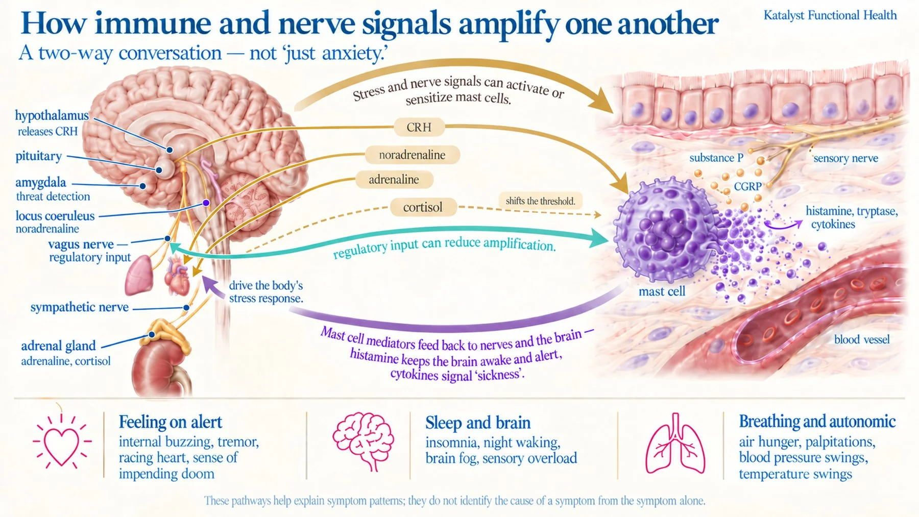
- Two speeds: the amygdala fires the locus coeruleus and sympathetic nerves in seconds, releasing noradrenaline right beside mast cells in tissue; the amygdala also signals the hypothalamus, starting the slower stress-hormone arm over minutes. CRH produced locally in tissue can act on mast cells directly. The fast arm is why a reaction can start before you have registered the stressor
- Stress → CRH → mast cells; mast cells → nerves → more stress signaling (Vanuytsel et al., 2014)
- Noradrenaline mainly shifts the threshold rather than firing the cell outright (mast cells carry both stimulating α and calming β2 adrenergic receptors)
- Sensitized nerves leak substance P, which degranulates mast cells with no immune trigger at all — someone can have florid reactivity with no allergen anywhere in the picture
- The absence of a safety signal is itself part of the problem: without it the cells stay more reactive, which is why nervous-system work is biology here and not a substitute for it
- Sleep loss lowers the threshold; poor sleep and flares feed each other
The cell changes itself
- In some inflammatory settings repeated triggering raises receptor numbers, so the baseline level of reactivity climbs and the threshold drops — though other conditions produce the opposite, and the cell becomes less responsive
- More mast cells recruited into inflamed tissue (SCF, NGF)
- Histamine, leukotrienes and IL-33 act back on the cell that made them
Gut chemistry
- Urease producers (H. pylori, Proteus, Klebsiella) and glutamine-fermenting bacteria raise luminal ammonia and pH
- Ammonia alkalinizes the mast cell interior, raises intracellular calcium and can release histamine without any immune trigger
- Mast cell granules must stay acidic to hold histamine; when that fails, histamine leaks and tryptase degrades
- Bacterial amines (putrescine, cadaverine) compete with DAO, so histamine clears more slowly even without touching the cell
- Aldehydes from yeast and fungal overgrowth load the same detox pathways histamine needs, so clearance bottlenecks while release is rising (more on ammonia and aldehydes in Neuroinflammation)
- Tissue pH shifts change how they behave in more than one direction, so an acidic environment doesn't simply calm them either
- Ammonia also injures epithelium, adding IL-33 and ATP
- This is a proposed gut mechanism drawn from laboratory work, not something established in people. We read it alongside your stool and organic acid picture rather than assuming it
Lost brakes
- Damaged epithelium can't stop making IL-33 and ATP
- Progesterone, vitamin D and omega-3 status shape stabilizing tone
- A separate question: is histamine also clearing slowly (DAO, HNMT)? Release and clearance are assessed separately
- Once mast cells have become dysregulated they can keep reacting even after the original trigger is removed — which is why recovery is management over time rather than one fix
- Chronic stress lowers vagal tone, weakening the body's own brake on mast cell release (Tracey, 2002; Rosas-Ballina et al., 2011) (see how immune and nerve signals amplify one another)
- Depleted glutathione → oxidative signals stay on
How immune and nerve signals amplify one another
Mast cells are wired into the threat circuitry. The amygdala reads danger, the hypothalamus starts the stress-hormone cascade, and adrenaline and noradrenaline flood the body. CRH made locally in tissue can act on mast cells directly. The mast cell's own mediators then feed back to the brain and confirm the threat.
Top-down and bottom-up
The loop above runs from the brain outward. It also runs the other way, and that direction explains more of how people actually feel.
The brain listening in
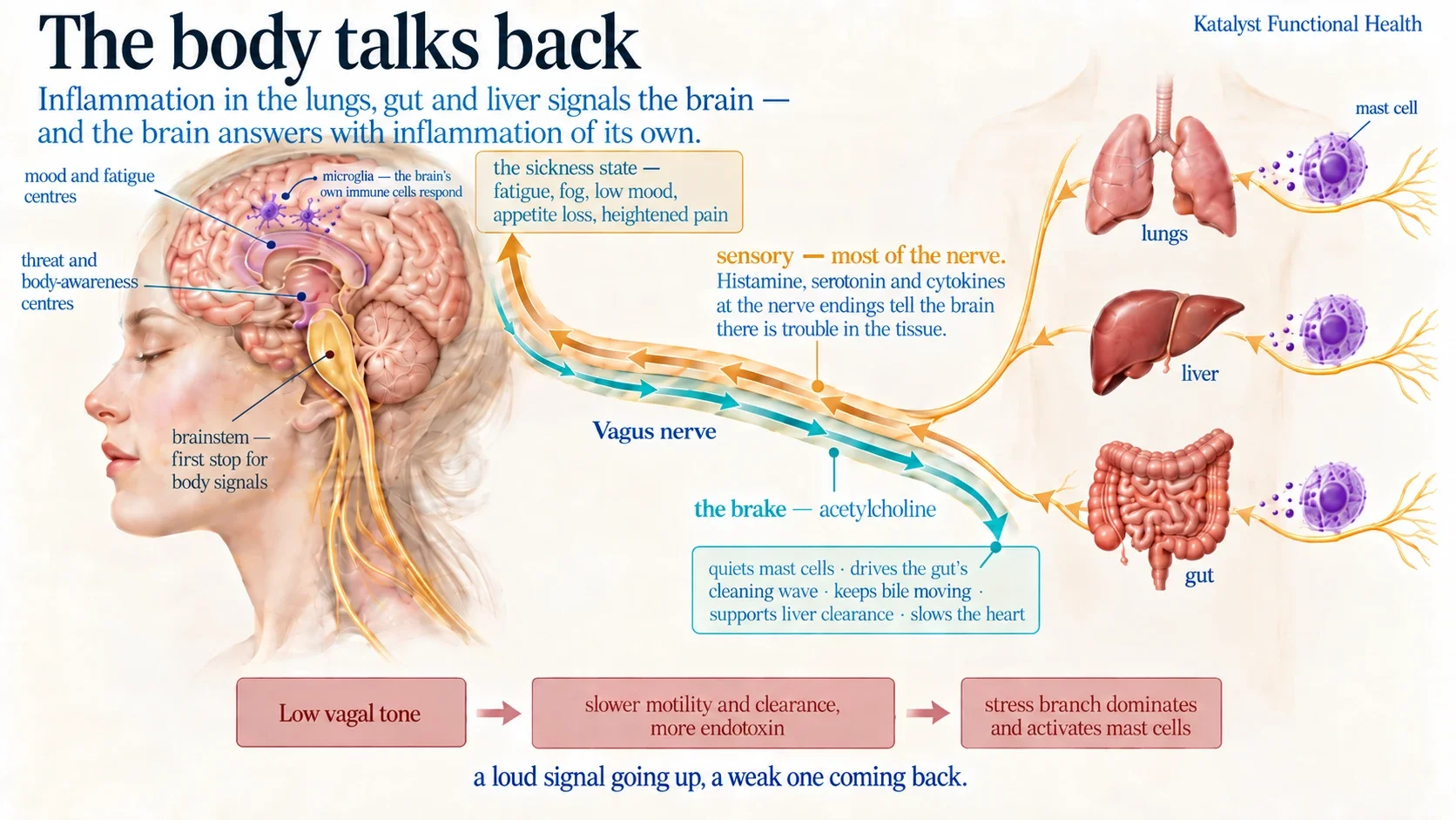
- Most vagus nerve fibers are sensory — they carry information up to the brain, not commands down
- Those sensory endings sit in the lungs, the gut and the liver — and mast cells sit right beside them in all three
- Histamine, serotonin and cytokines released there activate the nerve endings directly, so the brain is told there is a problem in the tissue before anything reaches it through the bloodstream
- That signal lands in the brainstem and travels on to the areas handling threat, body awareness and mood
- The brain then mounts its own inflammatory response: microglia, the brain's resident immune cells, switch on and produce cytokines locally. The message doesn't just get heard — it gets repeated inside the brain
- The result is the "sickness" state: fatigue, fog, low mood, appetite loss, heightened pain and a pulled-back feeling. It is a coordinated response to a message from the body, not a mood problem
- The hypothalamus sits in the middle of this, so hormone signaling shifts too. The clearest example is reproductive: inflammatory cytokines slow the pulse that drives the reproductive hormones, which is a recognized route to low testosterone, irregular cycles and reduced fertility during long illness. Thyroid and stress-hormone signaling shift in the same region for the same reason
Why the organ matters
- Lungs. Mold spores, fragments, endotoxin and particulates land on a surface dense in mast cells and richly supplied with vagal endings — one reason a damp building produces fatigue and fog, not only cough and congestion
- Gut. The largest mast cell population sits beside the largest sensory supply, which is why gut inflammation shows up as brain symptoms
- Liver. Reports immune status upward through the same reflex — so inflammation in tissue you cannot feel still registers centrally. The spleen sits on the receiving end: the vagus's calming signal reaches it through the splenic nerve
- The same nerve carries a brake back down. The best-established version of this reflex works through macrophages and a relay in the spleen; effects on mast cells themselves vary by tissue. Acetylcholine also does more than quiet immune cells. It drives the gut's cleaning wave between meals, keeps bile and digestive secretions moving, supports the liver's clearance work, and slows the heart
- So low vagal tone is not just a weak brake on mast cells. Motility slows and fermentation and endotoxin rise; bile and clearance slow; and with the calming branch quiet, the stress branch dominates — which activates mast cells from the other side. A loud signal going up, a weak one coming back, and a body tipped toward alarm
Why a flare can feel the way it does
These pathways help explain why immune and nervous-system symptoms interact. They are possible mechanisms, not a way to read the cause of a symptom from the symptom alone.
The sleep circuit runs both ways. Orexin neurons excite the histamine neurons of the tuberomammillary nucleus, and histamine at the H1 receptor is what keeps the cortex awake — orexin's arousal effect disappears in animals without the H1 receptor. The histamine neurons project back and densely innervate the orexin neurons, so the two drive each other. Histamine made in the body does not cross a healthy blood–brain barrier easily, but it still reaches this circuit: through sensory nerve signaling, cytokines and autonomic arousal, through a barrier that inflammation has loosened, and through mast cells that live in the brain and meninges. That is why lowering histamine in the body, not only in the brain, can improve sleep.
Mast cells and blood vessels
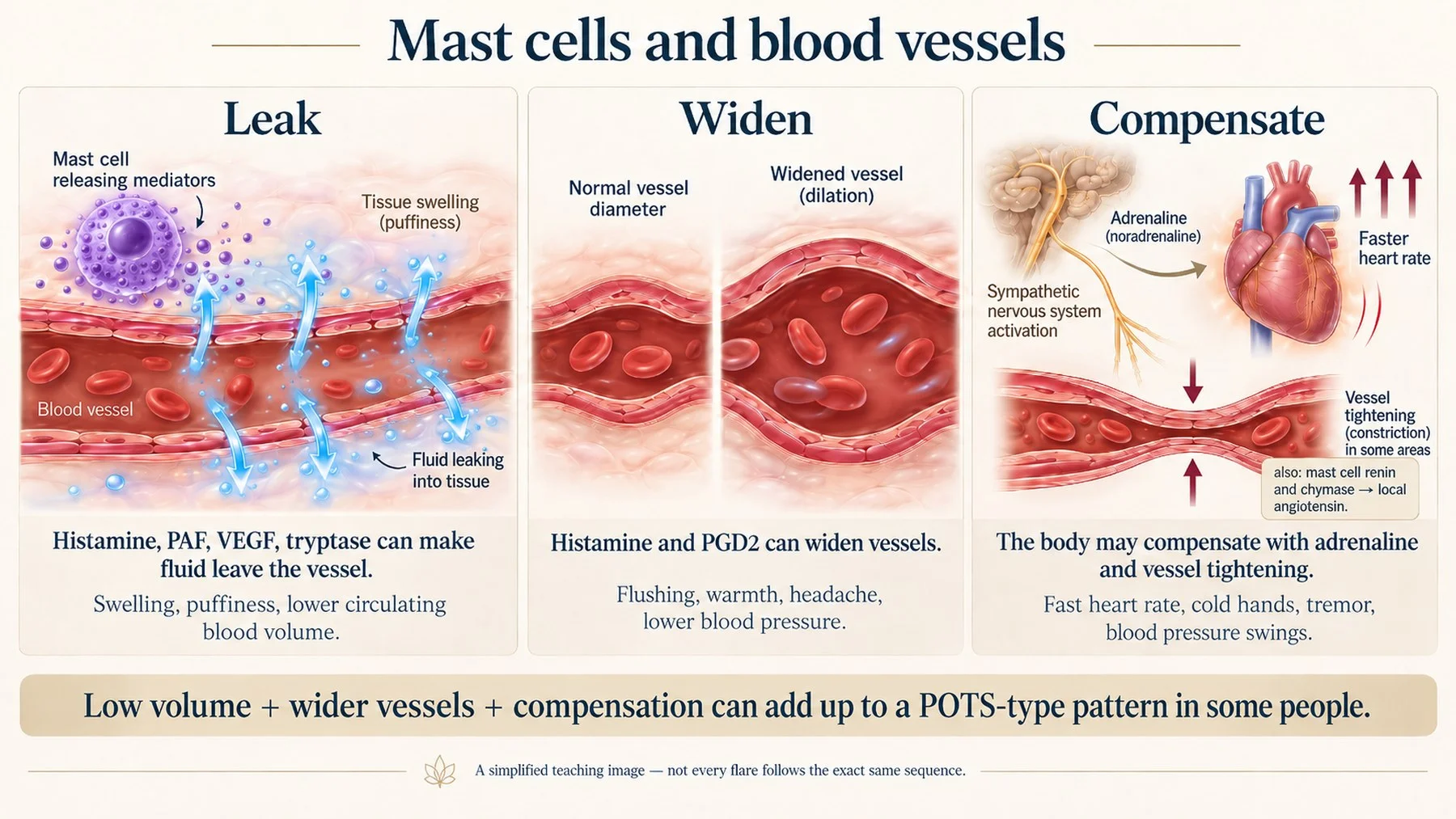
Mast cells sit on the outside of small blood vessels throughout the body. When they fire, the vessel leaks, widens, then the body slams on the brakes with adrenaline and angiotensin. In some people that push–pull can contribute to orthostatic intolerance and POTS-type symptoms — flushing, then cold hands, in the same hour.
Leak and widen
- Histamine, PAF, VEGF, tryptase open the vessel wall → fluid moves into tissue → swelling, puffiness, brain fog
- Fluid out of the vessel = less blood volume → low blood pressure on standing
- Histamine and PGD2 dilate vessels → flushing, warmth, headache
- Heparin activates factor XII, which makes bradykinin (Oschatz et al., 2011) → leakier vessels and swelling
Then constrict
- Low blood volume and falling pressure set off the body's reflex response: a sympathetic surge releasing noradrenaline and adrenaline → fast heart rate, cold hands, tremor
- Mast cells add to it locally — renin and chymase generate angiotensin II right in the tissue, which constricts vessels directly and amplifies noradrenaline release from nearby nerve endings
- Low volume + constricted vessels + a heart working harder to compensate can add up to a POTS-type pattern
Airway arm
- Histamine at the H1 receptor on airway smooth muscle tightens airways directly; leukotrienes C4/D4/E4 and PGD2 do the same and last longer, and thromboxane adds to it → air hunger
- Over-breathing drops CO2 → brain vessels narrow → lightheaded, tingling, foggy
Lymphatic arm
- Mast cells cluster along lymphatic vessels — in some tissues they associate with lymphatics more than with blood vessels
- Lymph doesn't flow passively. Collecting vessels pump, and histamine changes that rhythm: more frequent, weaker contractions, so the pumping is disorganized rather than simply slow
- Histamine, tryptase and VEGF make lymphatic walls leaky, so fluid that should be carried away seeps back into tissue — swelling that doesn't behave like ordinary fluid retention
- Mast cells also release VEGF-C and angiopoietins, which drive growth of new lymphatic vessels; in mastocytosis these track with symptom severity (Marcella et al., 2021)
- The consequence: waste, mediators and toxicants clear more slowly from tissue, which is one reason mobilizing anything too fast backfires
- MMP-9 and other enzymes stiffen the matrix the lymphatics run through, adding a second brake on flow
The vessel lining: the long game
Every vessel is lined with a sugar-protein "fur" called the glycocalyx. It helps the lining cells sense blood flow and produce nitric oxide, and keeps cells and LDL from sticking. Mast cell enzymes strip it — heparanase cuts the heparan sulfate chains that form its backbone, while tryptase, chymase, cathepsin G and MMP-9 break down the rest. Over years, sustained inflammation of the lining is a recognized vascular risk path.
Part of the inflammatory interplay
Plaque formation is a long, multi-factor process — lipid particles, blood pressure, blood sugar and insulin resistance, genetics and age all shape it. Mast cells aren't the whole story, but they aren't only a bystander either (Bot et al., 2015): they sit at the vessel wall adding inflammatory input, and they also help open the door upstream.
Endotoxin, or LPS (lipopolysaccharide), is part of the outer wall of gram-negative bacteria. In the gut it is normally kept on the right side of the barrier, but once it crosses into the blood it is one of the strongest inflammatory signals the immune system responds to. It shows how the loop closes. Gut mast cells release histamine and tryptase right at the intestinal lining, loosening the junctions between cells — so they help let endotoxin cross into the bloodstream in the first place. Once it is circulating, it binds the same TLR4 receptor on three cell types at once: on the vessel lining it switches on adhesion molecules and lowers nitric oxide; on circulating monocytes it drives the inflammatory response that follows them into the wall; and on mast cells it triggers more release. So mast cells appear at both ends — helping open the gut door, and responding at the vessel once the endotoxin arrives.
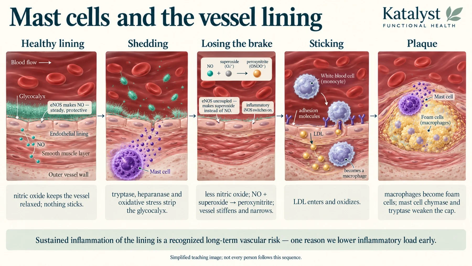
That sequence describes the large arteries, and it plays out over years. The same damaged lining has a second, faster consequence in the smallest vessels — and that one is usually what people are actually feeling.
When flow slows down
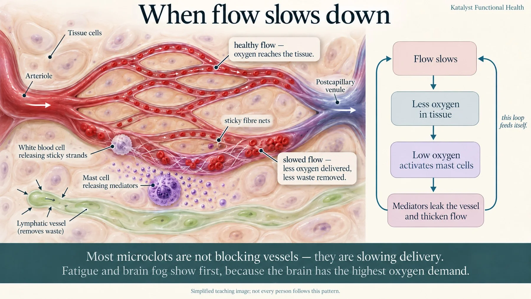
Capillaries are barely wider than a single red cell, so they feel a stripped lining and thickened blood long before a large artery does. Thick, sluggish blood there is not the same as a clot in a leg. It is a quieter problem: tissue gets less oxygen than it needs. Most microclots aren't blocking anything — they are slowing delivery.
The loop
- Slow flow means less oxygen in and less waste out
- Low oxygen is itself a mast cell trigger, so stasis activates the very cells that caused it
- Mediators loosen the vessel wall, fluid leaks into tissue, flow slows further
- Inflamed white cells arrive and throw out sticky fiber nets; platelets activate; small clots form that are harder to break down in an inflamed environment (Pretorius et al., 2021)
- Damaged lining cells release particles that trigger more clotting — the loop feeds itself
Where you feel it first
- The tissues with the highest energy demand feel it first: the brain as fatigue and brain fog, the heart as palpitations and exercise intolerance, often before anything shows on a standard test
- Liver and kidneys are the next highest-demand tissues
- Cold hands and feet, slow capillary refill, exercise intolerance
- Restoring flow alone isn't enough: while inflammation is high, mitochondria stay in an inefficient mode and can't use the oxygen that does arrive
Keeping this in proportion
- This is about the quality of flow through very small vessels, which is a different question from a clot in a large vein or artery
- It is measurable, starting with simple, widely available blood tests
- It responds to lowering inflammatory load, and to circulation and fibrinolytic support, started low and increased gradually
Why it belongs in a mast cell conversation
- Mast cell mediators make existing clotting tendencies worse, which is the one part of this that can become serious
- Fibrin is also the scaffold that biofilm-forming organisms build on, which links flow to persistent infection
Thick blood: three arms at once
Mast cells push the clotting system in three directions at the same time. Which one shows depends on the person and the moment.
- Thinning, straight away. Heparin boosts antithrombin, switching off key clotting factors; tryptase destroys fibrinogen (Prieto-García et al., 2012), chymase inactivates thrombin — the reason bruising, nosebleeds and heavy periods are common
- Clotting, within minutes. PAF is one of the most potent platelet activators known, and mast cells are a major source. Thromboxane clumps platelets and tightens vessels. Activated platelets then release their own serotonin, pulling in more platelets
- Clotting, over time. Cytokines drive the cascade, and damaged lining sheds particles that start coagulation
- The brakes fail at the same time. Protein C is switched on at the vessel lining by thrombomodulin, which inflammation turns down; protein S works alongside it. Antithrombin needs the heparan sulfate of the glycocalyx as its cofactor — exactly what has been stripped
- The end product is fibrin. The mesh that forms microclots, the scaffold biofilm-forming organisms build on, and a net that can trap circulating pathogens
Mast cells, hypermobility and dysautonomia
Three things travel together often enough that many people are eventually told they have all three: mast cell reactivity, dysautonomia such as POTS, and hypermobile joints. Not everyone with one has the others, and one did not simply cause the rest, but they are connected.
Connective tissue is the common ground. Mast cells live in it, and their enzymes (tryptase, chymase and MMP-9) remodel it. Where tissue is lax, veins stretch and blood pools on standing. Lax tissue also means more everyday strain, pain and micro-injury, and injured tissue releases the alarm signals that activate mast cells.
So the loop can run in any direction: mediators loosen tissue, loose tissue strains and signals, strain and low vagal tone activate mediators. It also means work on any one of the three tends to ease the others, which is the useful part.
Not all hypermobility is the same
It helps to separate four things that often get lumped together:
- Ehlers-Danlos syndromes (EDS). A growing spectrum of inherited connective tissue disorders. The 2017 international classification recognized 13 types linked to 19 genes (Malfait et al., 2017). More have been described since, including a new classical-like type in 2018 (Blackburn et al., 2018), and related overlap conditions continue to be defined. Every type but one is confirmed by finding the gene variant responsible
- Hypermobile EDS (hEDS). The most common type, and the exception: no causative gene has been confirmed, so it is still diagnosed on clinical criteria alone. A promising candidate is described below
- Hypermobility spectrum disorders (HSD). Symptomatic joint hypermobility that does not meet the criteria for hEDS. A separate diagnosis, not a milder form of EDS
- Acquired hypermobility. Not a formal diagnosis, but a pattern many clinicians describe: laxity that appears in someone who was never hypermobile, alongside inflammation, mast cell activation or chronic infection, and that may stabilize as those settle
What the research shows
The overlap is well documented (Kohno et al., 2021). In people with POTS and flushing, those with measurable mast cell activation had a distinct hyperadrenergic pattern: a sharp rise in heart rate on standing, and blood pressure that went up rather than staying flat (Shibao et al., 2005). Mast cell disorders are common in the Ehlers-Danlos syndromes (Seneviratne et al., 2017), and the hematologist Dr. Lawrence Afrin has proposed that some cases of hypermobile EDS may be rooted in mast cell activation itself (Afrin, 2021).
Two 2025 studies are changing how hypermobile EDS is viewed. The first compared blood proteins in 29 women with hypermobile EDS and 29 matched controls and found 35 that differed. Most were not structural: 80% belonged to immune, clotting or inflammatory pathways, and 43% to the complement system, the part of the immune system that tags and clears threats. Several complement proteins (C1q, C3, C8 and C9) were consistently low, and the cytokine pattern pointed to a dysregulated inflammatory response. The authors concluded that hypermobile EDS involves systemic immune dysregulation, not only a connective tissue problem (Griggs et al., 2025).
Why complement was low is not yet known. One possibility is consumption: in lupus, complement levels fall because ongoing immune activation uses them up, and an inherited lack of C1q is one of the strongest known risk factors for lupus. Whether something similar happens in hypermobile EDS has not been tested.
The second study looked at genes. In 200 people with hypermobile EDS, it found variants across the kallikrein genes, a family of enzymes that cut proteins, including a recurring KLK15 variant in several families. In mice, that variant altered tendons and heart valves and disturbed immune signaling. It is one of the most promising leads toward a gene for hypermobile EDS, though not yet confirmed (Gensemer et al., 2025). Together the two studies point the same way: hypermobile EDS involves immune signaling and protein-cutting enzymes, not only collagen.
Clinicians who see these patients don’t fully agree on how much mast cells drive the other two. Some, including cardiologist Dr. Andrew Maxwell, argue that mast cell enzymes can break down connective tissue enough to create hypermobility. Others, including mast cell specialist Dr. Tania Dempsey and pain specialist Dr. Linda Bluestein, see mast cell activation mainly worsening hypermobility, which can ease back toward a person’s baseline once it is controlled. Dr. Dempsey also sees POTS as a common consequence of mast cell activation. Pediatric cardiologist Dr. Jeffrey Boris is skeptical, because most of his patients were hypermobile before they became ill. What they agree on is that the three travel together and need to be looked at together.
How mast cells and inflammation can drive dysautonomia
Beyond the leaking, widening and tightening described in the blood vessel section, these are proposed mechanisms:
- Stretchier veins. Mast cell enzymes, including elastases and MMP-9, break down connective tissue in vein walls, so veins hold more blood on standing and less returns to the heart. Dr. Maxwell describes this as veins turning from a firm column into a saggy balloon. It remains a model rather than a proven cause
- Small nerve fibers. The small nerve fibers that carry autonomic signals to blood vessels and sweat glands can be damaged by inflammation. In 84 people with POTS, skin biopsies showed reduced small fibers in about 45%, and this tracked with reduced nerve supply to the heart (Haensch et al., 2014)
- The vagus nerve. Mediators released beside vagal sensory endings in the gut and lungs change what the brainstem hears, which shifts autonomic output (Berthoud & Neuhuber, 2000) (see how immune and nerve signals amplify one another)
- The upper neck. Lax ligaments at the top of the spine can strain the vagus nerve and slow drainage from the brain, a pattern described in the neck card in the next section
- Brain inflammation. The hypothalamus and brainstem run the autonomic system, and the same inflammation that causes brain fog can disturb them
What I see in practice
In my practice, I see acquired hypermobility regularly. Clients with no history of hypermobility develop it during a triggering event: mold exposure, a virus or other illness, or an infection. I see it the way I see most chronic illness: genetics can set the stage, but the environment pulls the trigger.
Two people can go through the same mold exposure or the same virus and come out with very different problems: joint laxity and POTS in one, gut or skin reactivity in another, brain fog or mood changes in a third. Which form the inflammation takes depends on each person’s history and total body burden: earlier infections, mold and toxin load, gut health, hormones, stress, past injuries and genetics. Reactive arthritis shows how the genetic piece can matter: after the same gut or urinary infection, people who carry the gene variant HLA-B27 are more likely to develop joint inflammation, and to have a more severe or lasting course (Colmegna et al., 2004). For hypermobility, I believe some people carry a predisposition to this inflammatory response and to the protease activation that loosens connective tissue. That last part is my clinical view, not an established finding.
A bigger problem since COVID
In a US database of more than 65 million adults, the rate of new POTS diagnoses rose sharply after March 2020, from about 1.4 to about 20 per million person-years, though some of that rise may reflect greater awareness and testing (Dulal et al., 2025). In a Cedars-Sinai cohort of nearly 285,000 people, the odds of a new POTS diagnosis were higher in the 90 days after COVID infection than in the 90 days before it (Kwan et al., 2022). In people with long COVID, mast cell activation symptoms were found to match those of people with diagnosed MCAS, even though their symptoms before COVID had looked the same as healthy controls (Weinstock et al., 2021). Hypermobility has not been tracked in the same way. Still, many functional practitioners who work in this area, myself included, now see far more mast cell activation, POTS and other dysautonomia, and hypermobility in their client populations than they did before the pandemic.
One explanation I find convincing, though it has not been tested directly, is that mast cells are a link in this chain. In the lab, the spike protein makes human mast cells release tryptase and chymase, the same protein-cutting enzymes that remodel connective tissue (Tsilioni & Theoharides, 2023). A small study found raised mast cell enzymes in the blood of people with long COVID (Wechsler et al., 2022). And in a UK study of about 3,000 people, those with hypermobile joints were about 30% more likely not to have recovered from COVID (Eccles et al., 2024). Put together, this suggests that in susceptible people the virus can tip mast cells into ongoing reactivity, and that reactivity may then drive both the autonomic symptoms and, in some, new or worsening joint laxity. That last step is what I see in practice.
One cell, many systems
Because mast cells are everywhere, chronic activation shows up as a pattern across systems rather than one disease. Mast cells do not operate in isolation: this map shows possible connections; it does not mean every pathway is active in you.
How I approach this
You cannot out-supplement a trigger that is still there. Stabilize symptoms while lowering the load, calming the wiring and repairing barriers — the levers run together.
1. Lower the load
Everything the immune system is still being asked to respond to. Usually several at once, not one.
- Environment. Where a water-damaged building is documented or the history points strongly to one, getting out of it is usually the single biggest lever (testing and next steps are in Happy Mold vs Angry Mold, Part II, and my own experience in Mold in a Newly Built Home). Also dampness, dust, fragrance, VOCs, new furnishings, renovation dust, pets if reactive
- Air where you sleep. The bedroom gets fixed first, because that is where repair happens
- Gut. Dysbiosis and endotoxin crossing a thin barrier; yeast, fungal and bacterial overgrowth; motility, so nothing sits and ferments
- Infections. Tick-borne, viral reactivation, parasites, sinus and oral colonization — worked in the right order and at the right pace, once terrain can tolerate it
- Toxicant burden. Mycotoxins, metals, pesticides, plastics; drainage open before anything mobilizing
- Food. Known allergies and identified sensitivities out. Temporary reduction of histamine, salicylate, oxalate or sulfur — whichever your pattern shows, and temporary is the operative word. Any restriction should be pattern-directed, time-limited and nutritionally supervised — a shrinking food list is a problem to solve, not a strategy. Also what comes with the food: organic where it counts to lower pesticide load, and glass or steel over plastic for storage, reheating and water
- Medication and supplement load. Excipients, dyes, fillers, and anything no longer earning its place
- Other inputs that stack. Alcohol, heat and cold extremes, overtraining, poor sleep
- EMFs. One of the cheapest levers here — reduce exposure, especially at night in the bedroom
2. Calm the wiring
Not a consolation prize. Nerve signals degranulate mast cells with no allergen involved, so this arm is biochemistry.
- Vagal tone. The nerve carrying the cholinergic brake to mast cells — breathing practice, humming, gargling, cold exposure if tolerated, gentle movement
- Limbic and safety signaling. The absence of a safety signal itself keeps the cells reactive. Whatever reliably tells your system it is safe counts — programs, somatic work, time in nature, familiar people
- Sleep. Protected first. Flares and poor sleep feed each other, and histamine is the brain's wake signal (Chikahisa et al., 2013; Huang et al., 2001)
- Breathing. Slower breathing holds CO2 and keeps blood flowing to the brain
- Pacing. Staying under the crash threshold; exertion is a trigger while flow and mitochondria are compromised
- Predictability. Routine, meal timing and light exposure lower the baseline alarm setting
3. Stabilize
Two separate questions: are the cells releasing too readily, and is what they release clearing too slowly?
- Flavonoids. Quercetin, luteolin, rutin. In practice these work more slowly than medication and reward patience, though direct comparisons are limited. Luteolin is the one with the most preclinical interest for neuroinflammation
- Botanicals. Nettle, perilla, chamomile, turmeric, holy basil, aloe, hops — introduced one at a time, small
- Vitamin C. Antihistamine in its own right and a cofactor for histamine breakdown. Plain ascorbic acid works and is the cheapest form; buffered or liposomal versions exist for people who react to the acidity, and whole-food sources like camu camu or rose hip are an option if a synthetic form doesn't sit well
- Clearance side. DAO with meals if gut breakdown is the bottleneck; the cofactors DAO and HNMT need — copper, B6, B5, magnesium, and intact methylation
- Stabilizing tone. Vitamin D (mast cells carry its receptor), omega-3s, and hormone balance
- Melatonin. Mast cells carry melatonin receptors, and in a flu model melatonin quieted mast cell activation through them. It also supports the sleep these cells disrupt. More in the melatonin section of Neuroinflammation
- Method. One change at a time, at the lowest sensible dose. If you react, the cause could be the active ingredient, the dose, an excipient, the formulation, the timing, or coincidence — worth working through rather than assuming
4. Repair the barriers
Damaged surfaces keep releasing the alarm signals that re-trigger mast cells, so repair is upstream work, not cosmetic.
- Gut lining. Tight junctions, mucus layer, secretory IgA — while the barrier leaks, endotoxin keeps arriving
- Microbiome. Rebuild rather than only remove; histamine-producing strains matter, so strain choice is not incidental. I name the main histamine-producing gut bacteria in Neuroinflammation
- Vessel lining and glycocalyx. Flavonoids, omega-3s, sulfur amino acids and nitric oxide support; circulation and fibrinolytic support
- Sinus and airway lining. Often the most overlooked surface. Where fungal colonization is suspected, it is worth assessing properly rather than assuming
- Mitochondria and antioxidant status. Glutathione and its precursors, B vitamins, minerals — paced to tolerance, since pushing detox too early is itself a trigger
- Connective tissue. Protein and amino acid sufficiency, vitamin C, gentle resistance work as tolerated
- Liver and bile flow. The liver carries out methylation-based histamine breakdown and a large share of toxicant processing, and bile movement carries processed toxicants out
- Nutrient repletion. Long illness depletes; low status keeps every brake weak
Curious how I work?
If you recognized yourself in these pages, the next step is not another list of supplements. It is working out what is loading your system, in what order to lift it, and what your body can tolerate right now. You can read how I put the pieces together in my approach, and it starts with a free 30-minute discovery call.
Selected references and further reading
Key studies and reviews behind the mechanisms described above. Many of the patterns in this article also come from clinical experience shared across practitioners, which doesn't always have a single paper behind it.
How mast cells activate and what they release
- Molderings GJ, Afrin LB. A survey of the currently known mast cell mediators with potential relevance for therapy of mast cell-induced symptoms. Naunyn Schmiedebergs Arch Pharmacol. 2023;396(11):2881-2891. PubMed 37243761
- Theoharides TC, Kempuraj D, Tagen M, Conti P, Kalogeromitros D. Differential release of mast cell mediators and the pathogenesis of inflammation. Immunol Rev. 2007;217:65-78. PubMed 17498052
- McNeil BD, Pundir P, Meeker S, et al. Identification of a mast-cell-specific receptor crucial for pseudo-allergic drug reactions. Nature. 2015;519(7542):237-241. PubMed 25517090
- Green DP, Limjunyawong N, Gour N, Pundir P, Dong X. A mast-cell-specific receptor mediates neurogenic inflammation and pain. Neuron. 2019;101(3):412-420.e3. PubMed 30686732
- Cao J, Papadopoulou N, Kempuraj D, et al. Human mast cells express corticotropin-releasing hormone (CRH) receptors and CRH leads to selective secretion of vascular endothelial growth factor. J Immunol. 2005;174(12):7665-7675. PubMed 15944267
- Allakhverdi Z, Smith DE, Comeau MR, Delespesse G. Cutting edge: The ST2 ligand IL-33 potently activates and drives maturation of human mast cells. J Immunol. 2007;179(4):2051-2054. PubMed 17675461
- Supajatura V, Ushio H, Nakao A, et al. Differential responses of mast cell Toll-like receptors 2 and 4 in allergy and innate immunity. J Clin Invest. 2002;109(10):1351-1359. PubMed 12021251
- Olynych TJ, Jakeman DL, Marshall JS. Fungal zymosan induces leukotriene production by human mast cells through a dectin-1-dependent mechanism. J Allergy Clin Immunol. 2006;118(4):837-843. PubMed 17030235
- Kempuraj D, Asadi S, Zhang B, et al. Mercury induces inflammatory mediator release from human mast cells. J Neuroinflammation. 2010;7:20. PubMed 20222982
- Kimata H. Enhancement of allergic skin wheal responses by microwave radiation from mobile phones in patients with atopic eczema/dermatitis syndrome. Int Arch Allergy Immunol. 2002;129(4):348-350. PubMed 12483040
- Tsilioni I, Theoharides TC. Recombinant SARS-CoV-2 spike protein stimulates secretion of chymase, tryptase, and IL-1β from human mast cells, augmented by IL-33. Int J Mol Sci. 2023;24(11):9487. PubMed 37298438
- de la Poza JFD, Parés AR, Aparicio-Calvente I, et al. Frequency of IgE antibody response to SARS-CoV-2 RBD protein across different disease severity COVID19 groups. Virol J. 2025;22(1):58. PubMed 40038712
- Riess T, Andersson SG, Lupas A, et al. Bartonella adhesin A mediates a proangiogenic host cell response. J Exp Med. 2004;200(10):1267-1278. PubMed 15534369
- Kempf VA, Lebiedziejewski M, Alitalo K, et al. Activation of hypoxia-inducible factor-1 in bacillary angiomatosis: evidence for a role of hypoxia-inducible factor-1 in bacterial infections. Circulation. 2005;111(8):1054-1062. PubMed 15723970
- Cayrol C, Duval A, Schmitt P, et al. Environmental allergens induce allergic inflammation through proteolytic maturation of IL-33. Nat Immunol. 2018;19(4):375-385. PubMed 29556000
- Zaitsu M, Narita S, Lambert KC, et al. Estradiol activates mast cells via a non-genomic estrogen receptor-alpha and calcium influx. Mol Immunol. 2007;44(8):1977-1985. PubMed 17084457
- Vasiadi M, Kempuraj D, Boucher W, Kalogeromitros D, Theoharides TC. Progesterone inhibits mast cell secretion. Int J Immunopathol Pharmacol. 2006;19(4):787-794. PubMed 17166400
- Mukai K, Tsai M, Starkl P, Marichal T, Galli SJ. IgE and mast cells in host defense against parasites and venoms. Semin Immunopathol. 2016;38(5):581-603. PubMed 27225312
Nervous system, brain and sleep
- Chikahisa S, Kodama T, Soya A, et al. Histamine from brain resident MAST cells promotes wakefulness and modulates behavioral states. PLoS One. 2013;8(10):e78434. PubMed 24205232
- Huang ZL, Qu WM, Li WD, et al. Arousal effect of orexin A depends on activation of the histaminergic system. Proc Natl Acad Sci U S A. 2001;98(17):9965-9970. PubMed 11493714
- Skaper SD, Facci L, Giusti P. Neuroinflammation, microglia and mast cells in the pathophysiology of neurocognitive disorders: a review. CNS Neurol Disord Drug Targets. 2014;13(10):1654-1666. PubMed 25470401
- Levy D, Burstein R, Kainz V, Jakubowski M, Strassman AM. Mast cell degranulation activates a pain pathway underlying migraine headache. Pain. 2007;130(1-2):166-176. PubMed 17459586
- Esposito P, Gheorghe D, Kandere K, et al. Acute stress increases permeability of the blood-brain-barrier through activation of brain mast cells. Brain Res. 2001;888(1):117-127. PubMed 11146058
- Tracey KJ. The inflammatory reflex. Nature. 2002;420(6917):853-859. PubMed 12490958
- Rosas-Ballina M, Olofsson PS, Ochani M, et al. Acetylcholine-synthesizing T cells relay neural signals in a vagus nerve circuit. Science. 2011;334(6052):98-101. PubMed 21921156
- Berthoud HR, Neuhuber WL. Functional and chemical anatomy of the afferent vagal system. Auton Neurosci. 2000;85(1-3):1-17. PubMed 11189015
- Forsythe P. Mast cells in neuroimmune interactions. Trends Neurosci. 2019;42(1):43-55. PubMed 30293752
- Nakamura Y, Nakano N, Ishimaru K, et al. Circadian regulation of allergic reactions by the mast cell clock in mice. J Allergy Clin Immunol. 2014;133(2):568-575. PubMed 24060274
- Vanuytsel T, van Wanrooy S, Vanheel H, et al. Psychological stress and corticotropin-releasing hormone increase intestinal permeability in humans by a mast cell-dependent mechanism. Gut. 2014;63(8):1293-1299. PubMed 24153250
Vessels, clotting and lymph
- Bot I, Shi GP, Kovanen PT. Mast cells as effectors in atherosclerosis. Arterioscler Thromb Vasc Biol. 2015;35(2):265-271. PubMed 25104798
- Oschatz C, Maas C, Lecher B, et al. Mast cells increase vascular permeability by heparin-initiated bradykinin formation in vivo. Immunity. 2011;34(2):258-268. PubMed 21349432
- Prieto-García A, Zheng D, Adachi R, et al. Mast cell restricted mouse and human tryptase·heparin complexes hinder thrombin-induced coagulation of plasma and the generation of fibrin by proteolytically destroying fibrinogen. J Biol Chem. 2012;287(11):7834-7844. PubMed 22235124
- Pretorius E, Vlok M, Venter C, et al. Persistent clotting protein pathology in Long COVID/Post-Acute Sequelae of COVID-19 (PASC) is accompanied by increased levels of antiplasmin. Cardiovasc Diabetol. 2021;20(1):172. PubMed 34425843
- Marcella S, Petraroli A, Braile M, et al. Vascular endothelial growth factors and angiopoietins as new players in mastocytosis. Clin Exp Med. 2021;21(3):415-427. PubMed 33687603
Allergy and IgE
- Reese TA, Liang HE, Tager AM, et al. Chitin induces accumulation in tissue of innate immune cells associated with allergy. Nature. 2007;447(7140):92-96. PubMed 17450126
- Gowthaman U, Chen JS, Zhang B, et al. Identification of a T follicular helper cell subset that drives anaphylactic IgE. Science. 2019;365(6456):eaaw6433. PubMed 31371561
Clinical picture
- Molderings GJ, Brettner S, Homann J, Afrin LB. Mast cell activation disease: a concise practical guide for diagnostic workup and therapeutic options. J Hematol Oncol. 2011;4:10. PubMed 21418662
- Seneviratne SL, Maitland A, Afrin L. Mast cell disorders in Ehlers-Danlos syndrome. Am J Med Genet C Semin Med Genet. 2017;175(1):226-236. PubMed 28261938
- Kohno R, Cannom DS, Olshansky B, et al. Mast cell activation disorder and postural orthostatic tachycardia syndrome: a clinical association. J Am Heart Assoc. 2021;10(17):e021002. PubMed 34398691
- Shibao C, Arzubiaga C, Roberts LJ, et al. Hyperadrenergic postural tachycardia syndrome in mast cell activation disorders. Hypertension. 2005;45(3):385-390. PubMed 15710782
- Afrin LB. Some cases of hypermobile Ehlers-Danlos syndrome may be rooted in mast cell activation syndrome. Am J Med Genet C Semin Med Genet. 2021;187(4):466-472. PubMed 34719842
- Griggs M, Daylor V, Petrucci T, et al. Proteomic discoveries in hypermobile Ehlers-Danlos syndrome reveal insights into disease pathophysiology. Immunohorizons. 2025;9(10):vlaf044. PubMed 40972649
- Haensch CA, Tosch M, Katona I, Weis J, Isenmann S. Small-fiber neuropathy with cardiac denervation in postural tachycardia syndrome. Muscle Nerve. 2014;50(6):956-961. PubMed 24647968
- Malfait F, Francomano C, Byers P, et al. The 2017 international classification of the Ehlers-Danlos syndromes. Am J Med Genet C Semin Med Genet. 2017;175(1):8-26. PubMed 28306229
- Blackburn PR, Xu Z, Tumelty KE, et al. Bi-allelic alterations in AEBP1 lead to defective collagen assembly and connective tissue structure resulting in a variant of Ehlers-Danlos syndrome. Am J Hum Genet. 2018;102(4):696-705. PubMed 29606302
- Gensemer C, Petrucci T, Beck T, et al. KLK15 alters connective tissues in hypermobile Ehlers-Danlos syndrome. iScience. 2025;28(9):113343. PubMed 40949095
- Colmegna I, Cuchacovich R, Espinoza LR. HLA-B27-associated reactive arthritis: pathogenetic and clinical considerations. Clin Microbiol Rev. 2004;17(2):348-369. PubMed 15084505
- Dulal D, Maraey A, Elsharnoby H, Chacko P, Grubb B. Impact of COVID-19 pandemic on the incidence and prevalence of postural orthostatic tachycardia syndrome. Eur Heart J Qual Care Clin Outcomes. 2025;11(5):698-704. PubMed 39775762
- Kwan AC, Ebinger JE, Wei J, et al. Apparent risks of postural orthostatic tachycardia syndrome diagnoses after COVID-19 vaccination and SARS-Cov-2 infection. Nat Cardiovasc Res. 2022;1(12):1187-1194. PubMed 37303827
- Weinstock LB, Brook JB, Walters AS, Goris A, Afrin LB, Molderings GJ. Mast cell activation symptoms are prevalent in Long-COVID. Int J Infect Dis. 2021;112:217-226. PubMed 34563706
- Wechsler JB, Butuci M, Wong A, Kamboj AP, Youngblood BA. Mast cell activation is associated with post-acute COVID-19 syndrome. Allergy. 2022;77(4):1288-1291. PubMed 34820848
- Eccles JA, Cadar D, Quadt L, et al. Is joint hypermobility linked to self-reported non-recovery from COVID-19? Case-control evidence from the British COVID Symptom Study Biobank. BMJ Public Health. 2024;2(1):e000478. PubMed 40018183